Constantly mutating painting by Japanese street artists for 1 week.
Monday, October 09, 2006
Labels: Constantly mutating painting by Japanese street artists for 1 week.
See More!
posted by Ammie @ 7:29 PM,
,
![]()
Women With Enlarged Breasts ‘Wanted’ By Police
 You can’t help but laugh after reading this story. The Police authorities in Berlin wouldn’t have thought that one day they will be handed an atypical “wanted” poster depicting enlarged naked breasts.
Well, the story goes like this. A German plastic surgeon who has been cheated out of payments by a number of women, has given the photos of their engorged breasts to the police. He thinks that the pictures may help the police to trace out those defaulter women.
He said that a lady named Tanja went outside after her $10,000 breast enlargement surgery and never came back. The cautious fellow now takes the amount in advance.
Labels: Women With Enlarged Breasts ‘Wanted’ By Police
See More!
posted by Ammie @ 5:22 PM,
,
![]()
Is He Pregnant...?
Wednesday, October 04, 2006
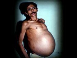
Doctors thought that Bhagat had a large tumor inside his stomach but were amazed when they found lots of bones, limbs, part of genitalia, some part of hair, jaws etc. This was just not what they were expecting. Inside his stomach was a half-formed creature which had developed feet and hands and had long fingernails.
It was then the doctors realized that creature found in Bhagat's stomach was no one but his own twin. He had one of the world's most bizarre medical conditions- fetus in fetu. This abnormality is extremely rare and occurs when a fetus gets trapped in its own twin and survives as a parasite and leaches its twin's blood supply.
Key words: fetus in fetu, spine of fetus, teratoma, twin, intraabdominal mass, parasitic twin.
The term fetus in fetu is used to point out an unequal division of totipotential cells of a blastocyst where the result is the inclusion of a small cellular mass in the more mature embryo. This is a form of monozygotic diamniotic twin pregnancy where the parasitic twin installs and grows in the body of its partner.1 This is a rare malformation that has some similarities with the retroperitoneal teratoma, but it is different from the latter by its fetiform aspect and the metameric segmentation of its spinal axis. The presence of a capsule covering this formation and a vascular pedicle is frequently encountered.2
In reality, one may find cases whose vertebral column is insufficiently calcified and therefore invisible on the plain radiograph. It may also be nonexistent but, because of the organogenetic differentiation, one may link this fetiform mass to the diagnosis of fetus in fetu.
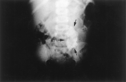 Fig. 1. Plain film of the abdomen. Calcified mass (arrows) at the left border of lumbosacral vertebrae L4, L5, and S1.
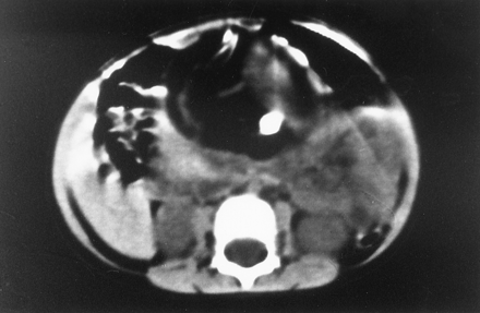 Fig. 2. Axial CT at the L2 level. In front of the vertebral column, there is a well-defined mass with 2 cystic formations. In the larger formation, which is anterior and on the left side, there are long and hyperdense opacities corresponding to fetal limbs.
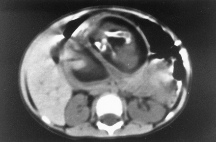 Fig. 3. Axial CT at the L3 level. In front of the vertebral column, there is a cystic mass containing fluid and some calcified opacities that correspond to fetal bones.
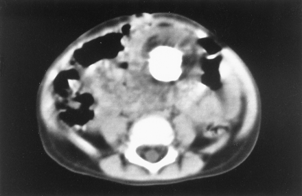
Fig. 4. Axial CT at the L3-L4 levels. In the midline of the abdomen, there is a round hyperdense opacity that corresponds to the fetal skull.
Based on these imaging findings, a diagnosis of fetus in fetu was thus made preoperatively.
The surgeon discovered a well-encapsulated retroperitoneal mass behind the transverse mesocolon. This mass had a pedicle that was connected to the superior mesenteric artery. Excision of the capsule revealed a yellowish fluid and an incompletely developed fetus covered by vernix caseosa.
The postoperative course was uneventful and the patient was discharged on the eighth postoperative day.
A radiograph of the specimen showed cranial bones and long bones but no vertebral column. On the macroscopic pathologic examination, the mass measured 20 × 8 × 5 cm and was composed of a head with hair, a trunk, and rudimentary limbs corresponding to an incompletely developed fetus. A soft vertebral axis was found behind the intestinal loops, colon, and liver. (Figs 5 and 6). Microscopically, there were hepatic cells, biliary tract cells, splenic tissue, and pulmonary tissue. Most of these cells were in blastic stage and not well-differentiated. The bony tissue was essentially composed of osteoblasts with little bone marrow. The nervous tissue was mostly formed by glial cells and not well-differentiated neurons.
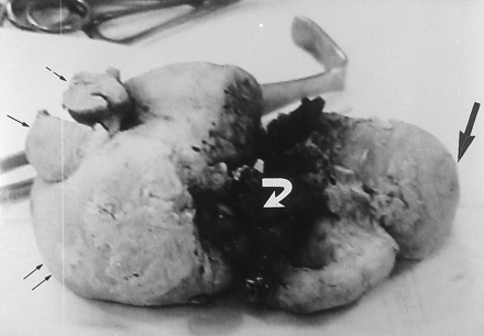
 Fig. 6. The postoperative specimen shows the fetus in prone position: the head on the left side (thick arrow) and the buttocks on the right side (small black arrows). The right shoulder and arm can be visualized (small white arrows) as a minor formation.
Watch A video...
Labels: Pregnant
See More!
posted by Ammie @ 5:44 PM,
,
![]()
JackAss New Game
Sunday, October 01, 2006
Labels: JackAss
See More!
posted by Ammie @ 11:28 PM,
,
![]()